Minimally Invasive Disc Surgery
Two operations take the pressure off a pinched nerve through a very small opening: an endoscopic discectomy, done through a portal about the width of a pencil with a camera, and a microdiscectomy, done through a small tube under an operating microscope. The four views below walk through both — where the incision actually goes, what the camera sees, what the surgeon sees down the microscope, and how little bone comes out.
The three incisions, on your back
All drawn to the same scale. Choose an approach below or tap an incision to explore it.
Endoscopic discectomy
~8 mmDMC Hub · Minimally invasive
Fragment removal through the foramen
Uniportal endoscopic discectomy · transforaminal route · L4–L5
Phases
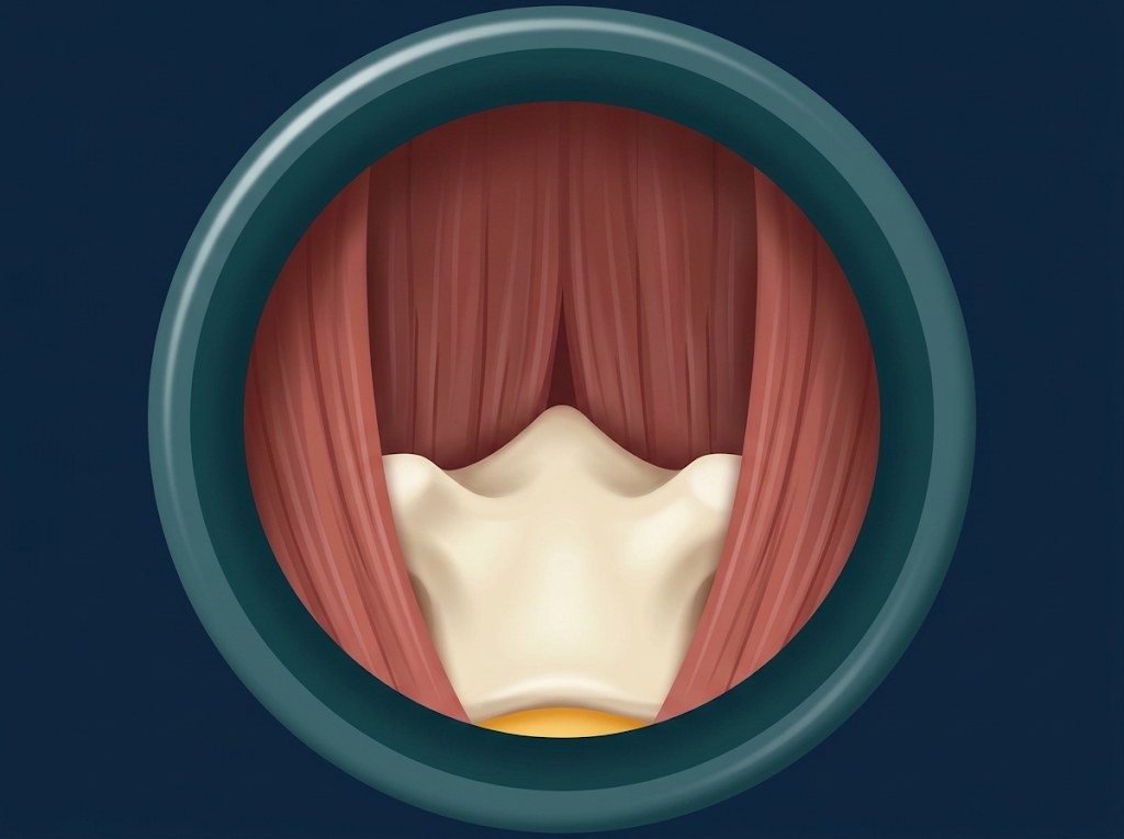
The window in the bone, seen from behind
How much of the bony roof each approach removes. Pick one to see the same two vertebrae before and after.
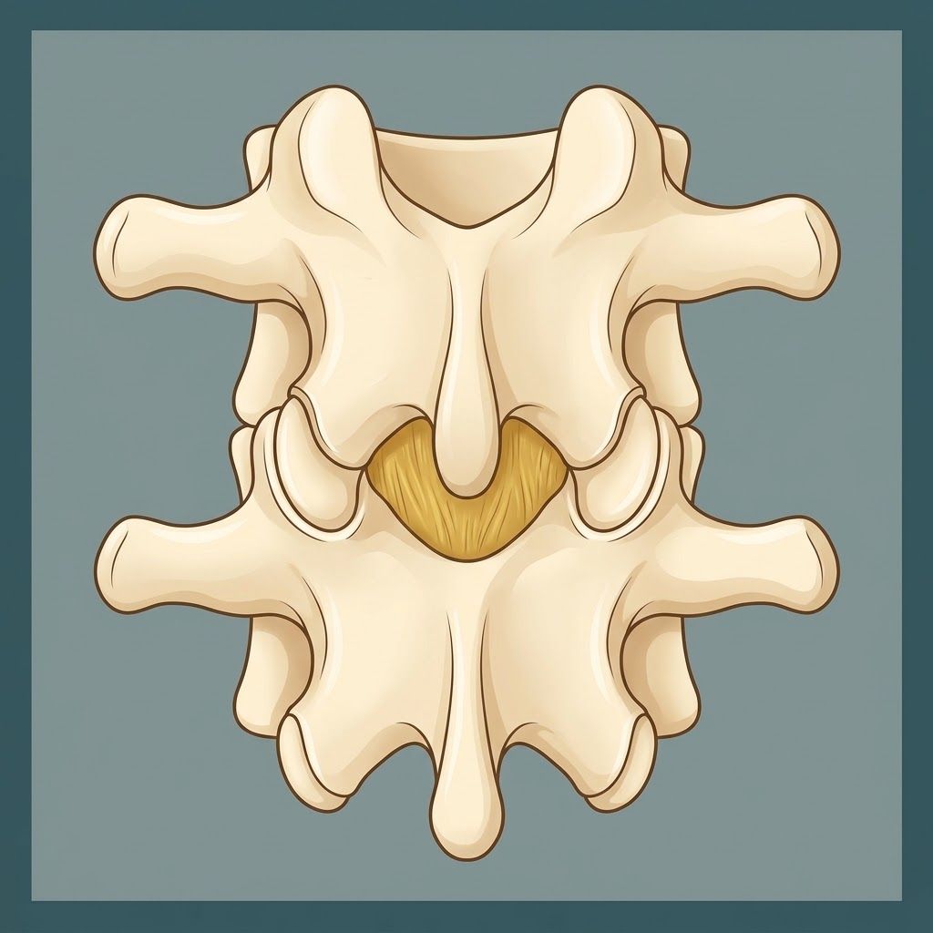
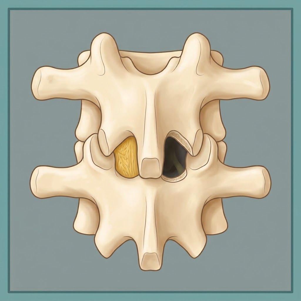
Microdiscectomy
~1 cm windowEducational tool only. These are simplified illustrations, not a recording of your own surgery. Which approach suits your herniation depends on your anatomy and is decided with your surgeon.